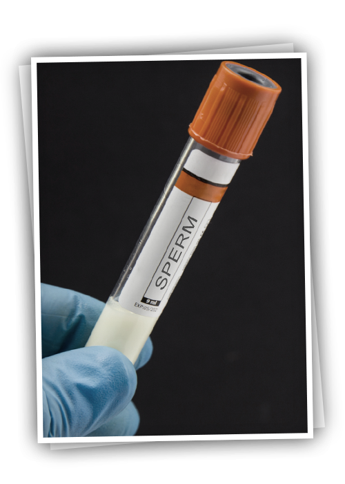
Micro-TESE
It is the process of looking for mature motile sperm by opening the ovary under anesthesia and taking samples from the canals using a microscope directly from the testis.
Contents
While the TESE process was first performed by taking more samples from the testicular tissue before the microscope was used, today less tissue from the correct foci is used by using the microscope and the process is called micro-TESE.
The success of the Micro-TESE procedure is higher than the TESE without the use of a microscope.
To Whom Is Micro-TESE Recommended?
Micro-TESE is recommended in case of azoospermia, which is thought to be due to insufficient sperm production in the testis.
Our goal in the micro-TESE process is to find spermatozoa, which are live, mature sperm cells with tails. Unfortunately, mature sperm cells may not be found in every surgery. Depending on the cause of azoospermia, the chance of finding spermatozoa in micro-TESE varies between 30% and 60%, and the success of conceiving varies between 30-40%. Although in vitro fertilization is performed with tailless sperm using the ROSI technique, which has been defined in recent years, this technique is still in the experimental stage.
How is Micro-TESE done?
Micro-TESE surgery can be performed with spinal (anesthetized from the waist) or general anesthesia. Additionally, local anesthesia can be applied. The testis is reached by making a 2-3 cm incision on the ovarian skin.
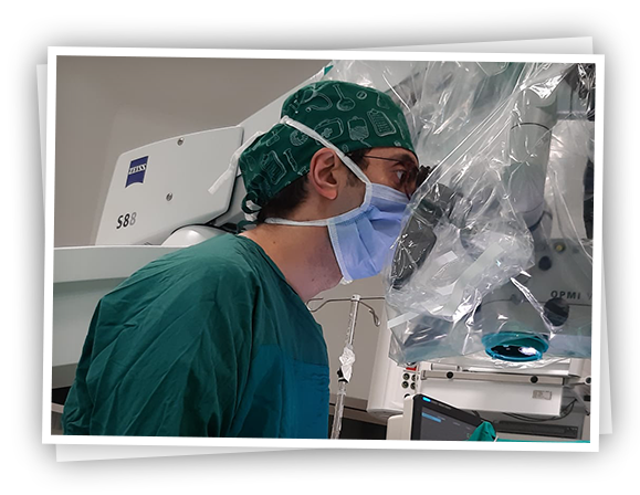
Using the operating room microscope, the testis is opened and the image is magnified 20-30 times and samples are taken from the full sperm producing-containing tubes. Samples taken are simultaneously examined by embryologists.
Sperm cells are investigated in the tubes. If enough mature motile sperm cells are found, these cells are used in IVF or frozen for later use. If sperm cannot be found, samples are taken from large tubes in the testicular tissue under the microscope and the sperm are investigated.
Samples are also taken for pathological examination in patients who cannot find sperm. If sperm is found, the testis and tissue layers are closed. If no sperm is found, the same procedure is applied for the other testicle.
Does the Micro-TESE Procedure Damage the Testicle? What Should Be Considered After?
It is a surgical procedure in which a small incision is made using a microscope and a microscope is used.
If it is done properly in experienced hands, the complication is very low.
Tissue damage to the testis is minimal since it is performed using a microscope.
Absorbable sutures are used, there is no need for suture removal. After a few days, you can take a bath. In order to prevent edema and swelling, it is recommended to wear tight-suspension pants for 1 week. Necessary drug treatments are given. Heavy exercise and sexual intercourse are not recommended for up to 2 weeks.
Should it be done again in those who do not have micro-TESE?
For those who could not find sperm in the first micro-TESE procedure, the patient can be re-evaluated with the pathology result and treated, and the procedure can be repeated 6 months later.
In cases where the first one was unsuccessful, the success rate was reported as 6-10%.
One of the factors affecting the success of the micro-TESE procedure is the genetic structure of the patient.
Patients should be evaluated beforehand in terms of chromosomal anomaly and gene deletions (AZFa-b-c) in the Y chromosome arm.
Again, the experience of the center, especially the embryologist and the surgeon, is very important.