Ureteral Stones | Uretero-Renoscopic Stone Crushing (URS)

Stones formed in the kidney may fall into the urinary canal, which we call the ureter, which carries urine from the kidney to the bladder. The stones sitting in the kidney may not cause complaints, but the stones falling into the urinary canal prevent the flow of urine and the urine accumulated behind causes swelling in the kidney. The kidney swells and the capsule with the outer neural network is stretched and stimulated.

Severe unbearable colic causes symptoms such as pain, nausea, vomiting, blood in the urine. Blood cells are seen in the urinalysis for diagnosis. On ultrasound, swelling in the kidney (hydronephrosis) is usually seen on the side of the stone. With tomography, the stone is clearly displayed.

In the treatment of ureteral stones, wait and see with drug therapy, stone breaking with sound waves and ureteroscopy with laser can be performed. The critical point is to apply the right treatment to the right patient.

İçindekiler

Which Stones Drop By Theyself?

The stone, which is thrown from the kidney to the ureter with urine, moves towards the bladder, that is, the urinary bladder, with the urinary pressure and peristaltic contraction of the ureter muscle. Many factors affect the progression-falling of stones thrown into the ureter:

  1. Stone size: Generally, we choose the treatment for ureteral stones by measuring the size of the stone. Small stones (less than 5mm) have an 85% chance of spontaneous passing. This ratio decreases as the size of the stone increases. For example, the 7mm stone has a 45% chance of falling, and the 10mm stone has a 25% chance of falling.
  2. Where the stone is located: There are 3 narrow regions of the ureter, which lies between the kidney and the urinary bladder. There is a possibility of getting stuck in these narrow areas. Stones in the upper part have a 22-48% chance of falling spontaneously, those in the middle have a 46-60% chance, and stones in the lower end have a 70-75% chance of falling.
  3. Duration of the stone staying in the same area: Stones waiting in the same place of the ureter for a long time cause edema and the ureteral wall swells and can prevent the stone from falling. Stones that stay in the same place for more than 30 days have a very low chance of falling.
  4. Surface structure of the stone: Irregular-surfaced-serrated-starlike stones have a lower chance of falling than smooth-surfaced stones.

Apart from these, the chance of stone passing is lower in patients with inflammation as a result of obstruction and in patients with severe swelling in the kidney. Again, child and female patients can pass their stones more easily.

These factors, alone or together, affect the falling of the stone. There may also be factors whose cause is not yet known. While waiting for the stone to pass by itself, kidney functions and the patient should be followed well.

If the patient presents to the emergency department every night due to severe pain, hinders his social activities and work life, and most importantly, if there is serious swelling and dysfunction in the kidney, he should not wait for the stone to pass any longer and start the treatment phase.

Drug therapy: Prostate drugs have been shown to be effective in lower-end ureteral stones of 5-7 mm in size in patients who are expected to pass the stone. Therefore, prostate drugs are given to these patients, even if they are women. Prostate drugs have the feature of expanding the urinary channels. It should be explained that male patients using prostate drugs will not have semen discharge after sexual intercourse and that this side effect is temporary. In addition to these drugs, patients with good kidney function can be given painkillers and edema solvents (non-steroidal anti-inflammatory). Patients with concomitant infections are also given antibiotic therapy.

ESWL treatment: For ureteral stones, the process of breaking the stones with external sound waves, namely ESWL, can be applied. This treatment is especially successful for 5-10mm stones in the upper ureter.

Uretero-Renoscopic
What is Stone Crushing (URS)?

It is the process of breaking up the stones in the urinary canal (ureter) and kidney with a thin endoscope with a camera (ureterorenoscope). The procedure is performed under anesthesia (general or spinal anesthesia) and completely naturally, without any incisions in the body.

In men and women, first, it is seen with a camera with endoscopes through the urethra, and it is moved inwards and the urinary bladder is reached. Then, the opening (orifice) of the duct that brings urine from the kidney is seen in the urinary bladder and a guide wire is sent here and this channel is entered. In this channel, it proceeds up to the kidney exit.

Stones at different levels in the canal can be seen directly. Stones in the kidney are reached with a flexible uretero-renoscope. Stones in the chambers (calyx) of the kidney can be easily reached and crushed with this device.

Are There Types of URS Surgery?

The naming is done according to the endoscope (ureteroscope) used during the surgery. If the ureteroscope used is a non-bending (rigid or semi-rigid) device, it is simply called URS. Rigid URS can only be used for stones in the ureter.

The surgery performed using a flexible endoscope (flexible ureterorenoscope) is called flexible URS (f-URS). Since angulation cannot be made with a rigid device, stones in the kidney or stones in the upper part of the ureter close to the kidney are reached using a flexible ureteroscope. In this process, stones are broken up using laser only.

How Are Stones Fragmented in URS Surgery?

The stones in the urinary canal are broken up with laser or pneumatic stone breaking tools. With the laser, the stones can be completely pulverized and these powders are excreted spontaneously in the urine. Larger stone pieces and devices called baskets are used in the urine.

Contact Us Pass

If the stones that remain in the urinary canal for a long time have caused serious edema on the wall of the canal, if the stone is very large, if the urinary canal is swollen due to obstruction, if there is an injury in the urinary canal during the surgery, a thin line is inserted into the urinary canal to ensure the flow of urine between the kidney and the bladder. tube (stent) can be placed.

In the flexible URS surgery applied for stones in the kidney, the stones are crushed by laser. A stent is usually placed after the procedure so that the stone fragments do not obstruct the urinary tract while pouring out of the urinary tract.

When is the Stent Placed During the Surgery Taken?

The stent can be removed after the stones broken in the surgery are removed from the urinary tract and the edema in the urinary tract is resolved. An X-ray or ultrasound may be done to make sure the stones are completely shed before stent removal.

The stent is usually removed 1 week to 3 weeks after surgery. The stent removal procedure is performed in the operating room under light anesthesia in the same way. After the stent is removed, patients can return home the same day.

The stents used (except for special stents remaining for a long time) are required to stay for a maximum of 3 months. Stents that remain in the body for a long time may cause problems such as urinary tract infection, bleeding, and petrification.

What kind of discomfort does the stent placed during the surgery cause?

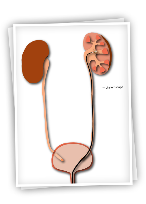
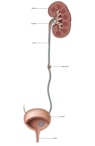
The stent is a thin plastic tube that provides the flow of urine between the kidney and the bladder and ensures the integrity of the urinary tract. They cause complaints such as flank pain, stinging, burning sensation, bleeding in the urine, frequent urination, and constant urination in patients.

The complaints caused by the stent in patients and their treatment methods have been the subject of many academic studies. Bladder relaxants (anti-muscarinics) and prostate drugs (alpha-blockers) can be given to relieve patients.

Who is the surgery recommended for?

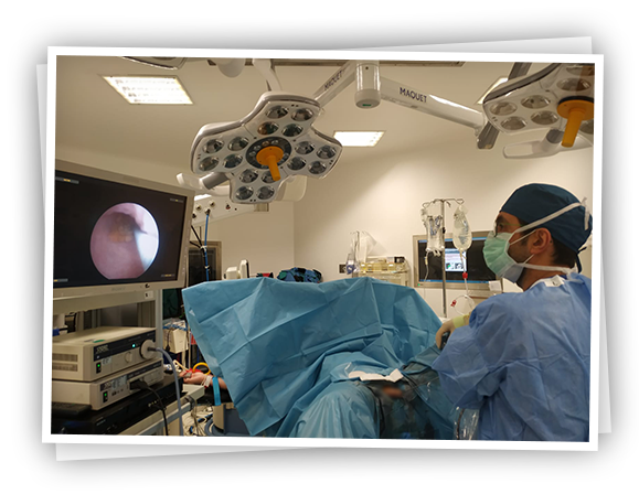
Patients with stones in the urinary tract (ureter) usually have 2 treatment options. External sound waves lithotripsy (ESWL) and ureteroscopic lithotripsy (URS). ESWL is recommended as the first treatment option for stones smaller than 1 cm in the upper part of the ureter, and URS surgery is recommended for stones larger than 1 cm. However, URS is also recommended for patients whose stones are not broken with ESWL.

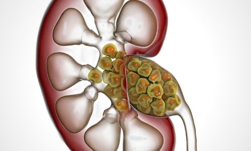
In the kidney, it is a particularly successful method for stones smaller than 2 cm. It can also be applied in larger stones, but several sessions may be required for complete stone-freeness.

Urs in Special Patients:

Since the URS procedure is performed through the urinary tract and without any incision, it can also be safely applied to those who use blood thinners.

Kidney-ureteral stones in overweight (obese) patients are difficult to treat. The success of sound wave lithotripsy (ESWL) in obese patients is lower than in non-obese patients. Percutaneous nephrolithotomy surgery is a very difficult and experienced procedure in these patients. f-URS is a safe treatment method in the treatment of kidney stones in obese patients.

Can URS Surgery be Applied in Children?

Since the urinary canals and kidneys of pediatric patients are much thinner and smaller than adults, the URS method can be used in pediatric ureter and kidney stones by using thinner and more sensitive endoscopes (ureteroscope). However, it is recommended to be applied especially in centers with experienced and sufficient technical equipment.

To Whom Is The Surgery Not Applied?

Since the procedure will be performed under general anesthesia, it cannot be applied to patients who are not suitable for anesthesia. In patients with urinary tract infection, the infection should be treated first and then the procedure should be applied.

How to Prepare Before the Procedure?

Before the procedure, the anesthesiologist will definitely want to examine you and see some of your laboratory tests, chest x-ray and heart electron. Before the surgery, your urine should also be checked for infection. If there is an infection, it must be treated beforehand. Again, the blood pressure, sugar or heart medications you use should be used in consultation with your doctor.

Eating and drinking water should be stopped 6-8 hours before the scheduled surgery time the night before the surgery.

Does URS Surgery Affect Sexual Life Negatively?

Since this surgery is performed through the urinary tract, men think that their sexual life will be adversely affected. In women, on the other hand, they think that their genitals may be damaged because the urinary tract is very close to the genitals. However, URS surgery does not harm sexual life or sexual organs of men and women.

What Are the Risks of URS Surgery?

urs ameliyatının riskleri - böbrek

Although the surgery is an endoscopic and incisionless procedure, it includes some risks. If the urinary tract is narrow enough to prevent the advancement of the endoscopes, balloon expansion can be performed. However, if it is still too narrow, the procedure can be postponed to the next session by placing a stent. The stents expand in the ureter and the second session (usually after 2 weeks) is successfully performed.

The most feared risk of the procedure is injury to the urinary tract during the procedure. This injury can range from a small tear in the ureteral wall to a rupture of the ureter's integrity. Minor injuries heal spontaneously after stent placement in the ureter, while complete ruptures are usually attempted to be repaired with open surgery.

Sometimes stones accompany the infection and this infection may not be detected with previous urinalysis. After the surgery, this infection can mix with the blood and cause serious conditions. In this case, it can be treated with appropriate antibiotics.

Apart from these situations, slight bleeding in the urine and pain on the operated side can be seen. A stone located in the upper part of the ureter may escape towards the kidney during crushing with URS. In this case, the stone in the kidney can be broken up with a flexible flexible ureterorenoscope.

How Many Days Will You Stay In The Hospital After The Surgery?

Patients can be discharged after hours after the operation, which is uneventful. However, patients are usually discharged 1 day after the procedure to make sure everything is going well. Patients who develop complications may need to stay in the hospital longer to complete their treatment.

How Long After To Return To Daily Activities Or Work?

The biggest advantage of endoscopic surgeries compared to open surgeries is the short return time to work and normal daily activities. It is recommended to spend the first few days after discharge, especially at rest. If severe pain, high fever and bleeding develop after the surgery, you should definitely consult your doctor.

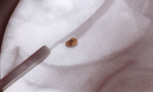
What to do with the removed stones?

The extracted stones are sent to the laboratory for analysis in order to learn the chemical substances they contain. Knowing the chemical content of the stone formed in the patient's kidneys can guide the doctors in preventing the recurrence of these stones.

In some patients, the stones become infected and contain germs (bacteria). During surgery, these bacteria can enter the blood and cause serious infections. For the most accurate and rapid treatment of these infections, the stones are placed in special containers during the surgery and sent to the microbiology laboratory in these patients. According to the results of this test, appropriate antibiotic therapy is given to the patients.

May Be Interested

Comments

Leave a Reply

Your email address will not be published. Required fields are marked *

TOP