Urinary Tract Stone Disease in Children – Dr. Tepeler

Although kidney stones are generally known as an adult disease among the people, unfortunately we encounter stone disease in all age groups in children. Its incidence varies in different geographies and has increased in recent years. It is more common in the climate zone, including our country, and in less developed countries.

With the widespread use of ultrasound, the frequency of kidney stones has increased. According to a study published in 2008, the incidence in school-age children in our country is 0.8%. If stone disease is not treated, it can cause organ loss and chronic kidney failure. Stone disease is the cause of 8% of children with chronic renal failure in our country. In the Southeast Anatolia region, which is the most common region, this rate rises to 20%.

İçindekiler

Which Children Are At Risk? Which Children Have Stones More Often?

  • Those with a family history of stone disease are particularly at risk.
  • It is more common in boys up to the age of ten and girls between the ages of 10-20.
  • Children who live in warmer areas and/or drink less
  • Those with congenital kidney-ureteral anomalies (such as ureteropelvic junction stenosis, ureterovesical junction stenosis, vesicoureteral reflux, neurogenic bladder)
  • Children with metabolic disease (idiopathic hypercalciuria, hypocitraturia, renal tubular acidosis, hyperuricosuria, enteric hyperoxaluria)
  • Genetic diseases (primary oxaluria, cystinuria, APRT deficiency, primary hyperoxaluria, renal tubular acidosis, Dent's disease) can be counted.
  • Children who are overweight, sedentary, especially those who are fed with heavy carbohydrates or protein, and consume sweetened beverages are also at risk.

What are the symptoms? What Complaints Does It Cause?

Stone disease can progress in a very different clinical picture in children. While kidney stones are the first diagnosis that comes to mind in severe pain in adults, clinical symptoms in children can be very different. Stone disease can be detected even in children who have no complaints. However, stone disease should be suspected in children with abdominal pain, restlessness, nausea, vomiting, burning and bleeding in the urine, frequent urination and growth retardation.

How is Kidney Stone Diagnosed in Children?

In pediatric patients, the diagnosis of kidney stones is usually made at the end of the process that starts with the detection of blood in the urine as a result of the tests performed by the pediatricians. As a result of the urinalysis, blood cells (erythrocytes and leukocytes) or crystals belonging to the stone are seen. Kidney functions and infection values (CRP, urine culture) are checked. Definitive diagnosis is made by imaging methods. For this purpose, ultrasound is most often used. Ultrasound is the safest method as X-ray is not used.

Contact Us Pass

With ultrasound, the location of the stone, its size, and the degree of swelling in the kidney are learned. X-ray film is also one of the methods that can be used in follow-ups. Low-dose radiation tomography can be used in cases where ultrasound is insufficient or in patients who are considered for surgery.

What are the Treatment Methods?

Erişkinlerde olduğu gibi çocuklarda da taşın idrar yollarında bulunduğu yer (böbrek içi, idrar kanalı veya mesane), taşın boyutu, sertlik derecesi, böbreklerde tıkanma varsa derecesi, böbrek fonksiyonları tedavi kararını ve tedavi çeşidini belirlemede önemlidir. Bütün tedavilerde öncelikle varsa enfeksiyon tedavi edilmelidir.

Kidney stones: We basically prefer monitoring, stone breaking, flexible ureteroscopic laser therapy and percutaneous nephrolithotomy methods in the treatment of kidney stones. Children have the same organs as adults, but because they are much smaller and sensitive in size, more miniaturized (thinner in small diameters) surgical instruments are used.

Follow-up: It is mostly applied for stones less than 1 cm in the kidney chambers that do not cause any complaints and do not obstruct the urinary canals. However, ultrasound controls should be performed at intervals of 3-6 months due to the risk of the stones growing or falling into the urinary canal. In studies conducted with adults, 25% of the patients definitely need treatment during the follow-up period. Metabolic evaluation for the underlying metabolic problem must be performed. If any, treatment should be given for the treatment of the detected metabolic problem.

Stone breaking with sound wave (ESWL): ESWL, which is a treatment method frequently applied in adults, can also be applied in pediatric patients in experienced centers.

It is based on the breakdown of the stone in the kidney by focusing the shock waves produced in a generator. The broken stone is then expelled from the urinary canal with the urine. Its success depends on many factors such as the hardness of the stone, its location in the kidney, and the size of the stone. It is especially applied for stones smaller than 2 cm. The disadvantages are that it requires additional sessions and requires anesthesia in young children.

Laser therapy with flexible ureteroscopy (retrograde intrarenal surgery):

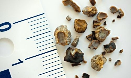
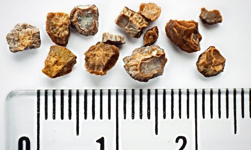
Today, thanks to the development of much thinner endoscopes (devices with cameras that show inside the body), kidney stones are treated in pediatric patients without making any incisions. It is the process of entering the external urinary canal with endoscopes, going up to the kidney, breaking the stone into powder with laser, and removing the small pieces with a basket. It is the ideal treatment option especially for stones of 1-2 cm in size.

In children with narrow ureters, a stent is inserted in the first session to expand the ureter (urinary canal between kidney and bladder). In the second session, which will be held 2 weeks later, the transition to the kidney will be easier. Stent placement can be done after the stones are broken up with laser, depending on the surgeon's decision.

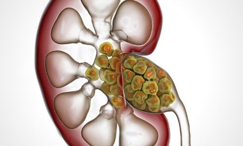
Percutaneous stone surgery (Percutaneous nephrolithotomy - PNL):

This method was first applied to adults in 1979, and it was successfully performed in children in 1995-2000 with the development of small-diameter endoscopes. It is the process of breaking up and removing the stones in the kidney by creating an entrance way of approximately 6-10 mm from the skin to the kidney from the patient's back area under anesthesia. In this way, no stones remain in the kidney. The success rate is higher than other treatment methods, especially in stones larger than 2 cm.

The risks of the surgical technique have decreased with the introduction of thinner-diameter instruments and especially the laser. With mini (miniperc), ultramini (UMP) or micro percutaneous nephrolithotomy (microperc) methods, the procedure can be performed successfully, especially in children under 1 year old. I have articles in which I apply microperc and ultramini methods to many of my patients and share my experiences on these issues in the international academic community.

Ureteral stones:

Although the diameter of the ureter is thinner in children, the ability to stretch is greater. Pediatric patients can pass ureteral stones more easily than adults.

Medication:

Especially small (less than 4 mm) stones have a higher chance of spontaneous passing. As their size increases, the chance of falling decreases or they cause more complaints when falling. Symptomatic treatment (relieving complaints) is usually given as drug therapy. Children with pain can be given painkillers and, if there is an infection, treatment for it. Prostate drugs used in adults were found to be particularly effective for stones of 4-10 mm in size located at the lower end of the ureter. Although it is not yet included in urology guidelines, it is a treatment option with proven effectiveness in suitable patients.

Shock wave therapy (ESWL):

Stone breaking with extracorporeal shock waves is applied in ureter (urinary tract) stones as well as in kidney stones. It can be applied to ureteral stones of 4-10 mm in size, especially in the upper part, which do not benefit from drug therapy. In pediatric patients, the procedure is performed under anesthesia. However, it does not require staying in the hospital or the applied center. The hardness and size of the stone, the experience of the center are the factors affecting the success.

Stone treatment with endoscopic laser:

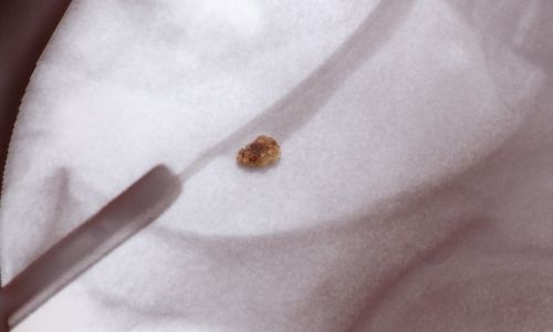
Under anesthesia, the patient's urethra is visualized with thin endoscopes, and firstly the bladder (urinary bladder) and then into the ureter, the stone is reached and the stone is completely pulverized with laser. A sample is definitely taken for stone analysis. If the stone has edema in the ureter wall or depending on the surgeon's decision, a stent is placed in the ureter at the end of the operation. Pediatric patients are usually hospitalized and followed up for at least 1 day. The placed stent is removed after 2 weeks. After the procedure, children may bleed in the urine for a few days.

May Be Interested

Comments

Leave a Reply

Your email address will not be published. Required fields are marked *

TOP